How Is Neuropathy Measured? Understanding What the Tests Tell Us
Peripheral neuropathy can cause numbness, tingling, burning, pain, weakness, balance problems, and unusual hot or cold sensations. But symptoms alone do not always tell us how much nerve function has been affected. Someone can have considerable sensory loss with very little pain, while another person may experience significant burning or tingling even though the area of sensory loss is relatively small.
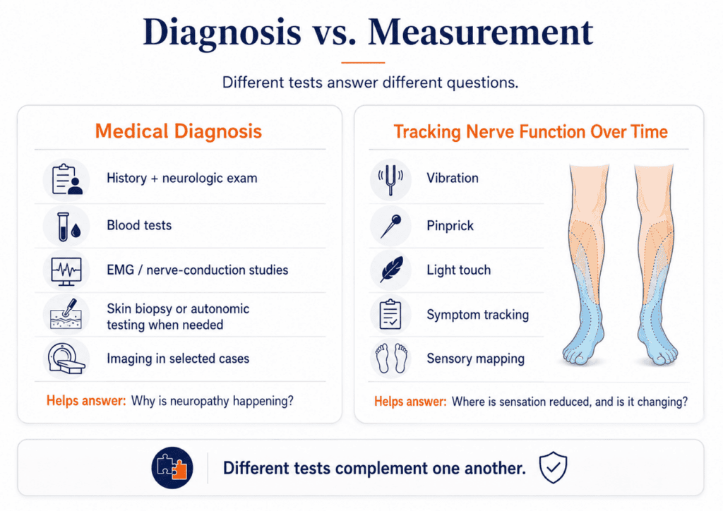
That is why evaluating neuropathy usually involves more than asking how your feet feel. Healthcare providers use a combination of your medical history, physical examination, diagnostic testing, and sensory testing to answer two related questions: Why is neuropathy occurring, and how much nerve function has been affected?
Diagnosing Peripheral Neuropathy
A neuropathy evaluation usually begins with a medical history and neurologic examination. Your provider may ask when symptoms started, whether they affect both sides equally, whether you have numbness, pain, weakness, or balance problems, and whether you have conditions or exposures known to affect nerves. Diabetes, vitamin deficiencies, thyroid disease, kidney or liver problems, certain medications and chemotherapy drugs, alcohol use, toxins, and inherited conditions are among the many possible contributors.
The neurologic examination may include sensation, reflexes, strength, gait, coordination, and balance. The pattern of symptoms is important. Problems affecting both feet in a relatively symmetrical “stocking” distribution suggest a different process than symptoms limited to one nerve, one side of the body, or a specific nerve root.
Blood testing is also commonly used to look for an underlying cause. Depending on the patient, testing may include blood glucose, vitamin B12, thyroid function, blood counts, kidney and liver function, and testing for abnormal proteins. Additional laboratory tests can be ordered when the history or examination points toward a particular cause.
Some patients also undergo nerve-conduction studies and electromyography (EMG). These tests measure how electrical signals travel through nerves and muscles and can help determine whether a problem involves peripheral nerves, nerve roots, muscles, or another part of the neuromuscular system. They are particularly useful when symptoms are unusual, asymmetric, rapidly progressive, or accompanied by significant weakness.
Standard nerve-conduction studies primarily evaluate larger nerve fibers, however, so they do not identify every type of neuropathy. When small-fiber neuropathy is suspected, specialized testing such as skin biopsy or autonomic testing may sometimes be appropriate. Imaging such as MRI is generally reserved for situations where the examination suggests a structural problem such as spinal stenosis, a herniated disc, or nerve compression.
The important point is that there is no single test that tells the entire story of neuropathy. Different tests answer different questions.
Measuring Sensory Function
Once neuropathy has been identified or is strongly suspected, another question becomes important: Where is sensation reduced, how extensive is the affected area, and is it changing over time?
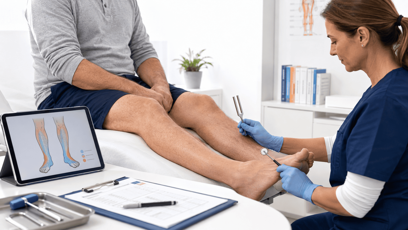
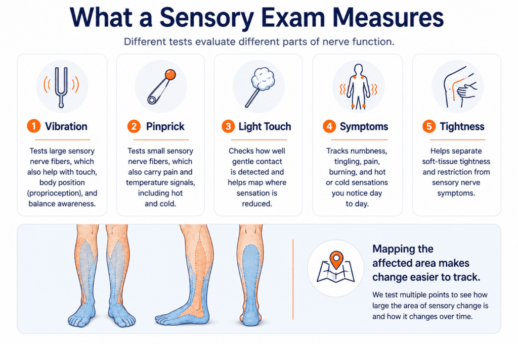
A sensory examination can evaluate several different types of nerve function. Vibration is often tested with a tuning fork. Pinprick testing evaluates how clearly the nervous system recognizes a small, controlled sharp stimulus. Light-touch testing measures the ability to recognize gentle contact with the skin. These tests are performed at multiple locations because neuropathy typically affects a pattern rather than one isolated spot.
Symptoms are also important. Numbness, tingling, burning, shooting sensations, pain, temperature changes, and tightness can all provide useful information. What the patient reports and what can be measured objectively should be considered together.
One particularly useful concept is sensory mapping. In the common distal symmetrical form of peripheral neuropathy, sensory loss typically begins at the ends of the longest nerves—often the toes and feet—and may gradually progress upward toward the ankles and lower legs. By testing a series of locations, the affected surface area can be mapped rather than simply labeling a patient as having “neuropathy.”
That gives clinicians something that can be compared over time.
What Does Improvement Look Like?
Neuropathy improvement is often gradual, which can make it difficult to recognize from one day to the next. A patient might still say, “My feet feel numb,” even though measurable sensory function has already changed elsewhere.
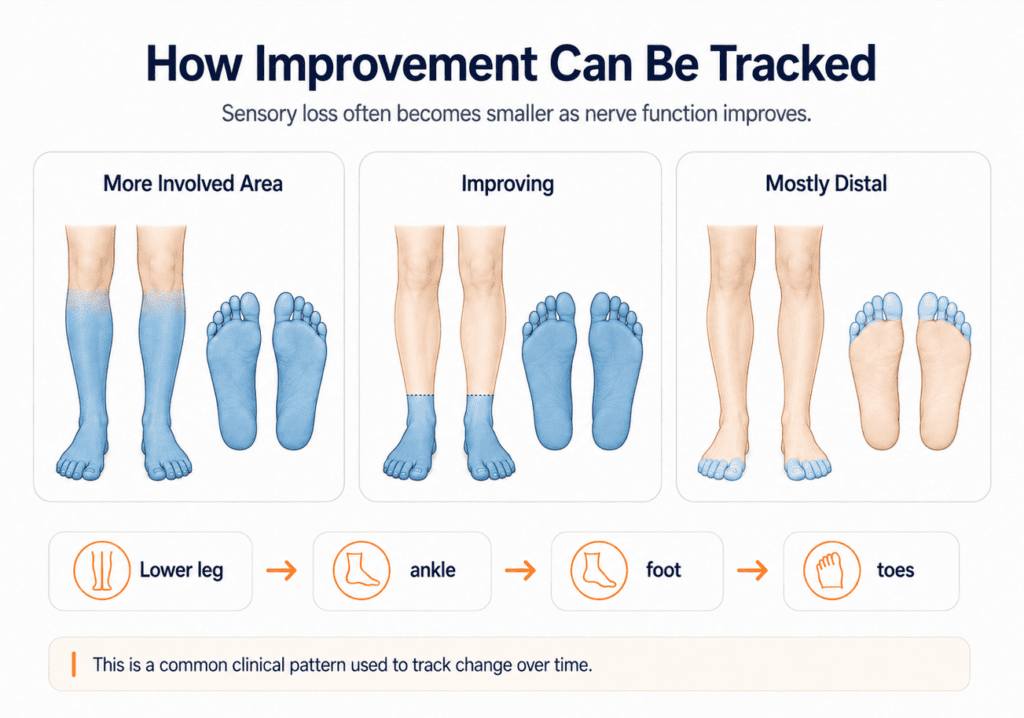
In our clinical experience at Realief, when sensory function improves, the affected area frequently shrinks from the top down, opposite the direction in which sensory loss typically progressed. For example, someone whose sensory abnormalities initially extended from the feet nearly to the knees may first show improvement higher on the lower leg. Over time, the remaining abnormal area may move progressively downward toward the ankle and foot.
As improvement reaches the foot, the arch is often one of the first areas on the bottom of the foot where change becomes apparent. The toes and the area where the toes meet the ball of the foot are often among the last places where sensory abnormalities remain.
That does not mean every patient follows exactly the same pattern or timeline. Rather, it gives us a recognizable clinical pattern that can help us interpret repeated examinations.
This is also why a patient may not immediately notice early improvement. Areas higher on the leg may have had only mild sensory loss and may never have caused noticeable symptoms. A clinician may therefore measure improved vibration or pinprick sensation before the patient notices a major change in the areas that bother them most.
Why Re-Examination Matters
Repeated examination gives neuropathy care a baseline and a way to measure change. Instead of relying only on whether someone feels better on a particular day, sensory findings can be compared with the original examination.
That distinction matters because symptoms can fluctuate. Tingling may temporarily increase. Tight tissues that were not very noticeable when sensation was reduced may become more apparent as sensory function improves. Pain can improve at a different rate than numbness. Objective testing helps place those experiences in context.
At Realief Neuropathy Centers of Minnesota, sensory mapping and repeated examination are used to track how the affected area changes during care. This does not replace the medical diagnostic workup used to determine why neuropathy developed. Many patients have already gone through that process before they reach us. Our focus is on measuring sensory function over time so that treatment decisions are based on more than symptoms alone.
“Testing for neuropathy” can mean several different things. A medical evaluation may help determine whether neuropathy is present and what may be causing it. Blood tests, EMG and nerve-conduction studies, skin biopsy, autonomic testing, or imaging may all have a role depending on the individual situation.
Sensory testing answers a different question: How well are the nerves functioning in specific areas, and is that changing?
Neither approach replaces the other. A blood test cannot tell you how large an area of numbness is. A tuning fork cannot tell you whether vitamin B12 deficiency is causing neuropathy. An EMG cannot fully evaluate every small nerve fiber.
Different tests provide different pieces of the picture.
And when neuropathy is being followed over time, one of the most useful questions is not simply, “How do you feel today?”
It is also:
“What has actually changed since we started?”
About the Author
Dr. Timothy Kelm DC is the founder of Realief Neuropathy Centers of Minnesota and has spent over 20 years focused exclusively on the evaluation and treatment of peripheral neuropathy. He has worked with thousands of neuropathy patients and delivered tens of thousands of neuropathy-focused treatments.
He is associated with published clinical research conducted in collaboration with the University of Minnesota and holds a nationally issued patent related to neuropathy treatment methodology. He has delivered public educational presentations and trained physicians nationally on structured neuropathy care.
Over 20 years ago, his interest in neuropathy began after repeatedly seeing patients who were told there were no good options. He believed then – and continues to believe today – that neuropathy should not define your life.
References
Hamblin MR. Photobiomodulation for peripheral nerve injury. Photomed Laser Surg.